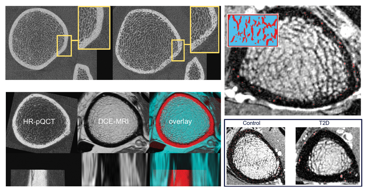
A decade ago, Thomas Link, MD, PhD, was senior author on a paper in J Bone Miner Res1 that suggested that severe deficits in cortical bone quality are responsible for fragility fractures in postmenopausal diabetic women. Building on that work, our Bone Quality Research Lab has developed a technique to visualize intra-cortical vessels and assess the structural changes that can degrade bone strength.
To understand how the normal vascular structure is altered, rendering the compact cortical shell very porous in some patients, our lab developed a technique that uses both dynamic contrast-enhanced MRI and high-resolution peripheral quantitative computed tomography (HR-pQCT)2. Using this new capability, we have analyzed baseline data of the distribution and size of intracortical vessels in patients with diabetes compared to healthy controls.
One imaging challenge is deducing which…


