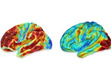
 Normal aging leads to frontocortical atrophy. The degree to which this complicates the use of frontotemporal atrophy as a diagnostic criterion for the frontotemporal dementias (FTDs) has not been reported. The present case-control study compared frontotemporal volumes delineated with semi-automatic brain region extraction [n=30 controls vs. 16 behavioral variant FTD (bvFTD) vs. 14 primary progressive aphasia]. Logistic regression identified those regions least helpful for distinguishing bvFTD and primary progressive aphasia from controls. Linear regression tested the correlation of duration of illness to atrophy severity. The control group showed high variance in volumes. Controls had right frontal lobe volumes that overlapped considerably with bvFTD volumes, but, as anticipated, the left anterior temporal volumes of interest showed 91% accuracy in distinguishing the aphasic subgroup…
Home Alzheimer's Research Overlap in Frontotemporal Atrophy Between Normal Aging and Patients With Frontotemporal Dementias


