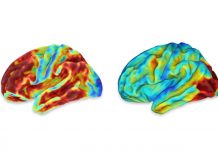
We found that sCoV increased between SCD, MCI and early probable AD groups in GM and in the temporal lobe. As expected, sCoV was negatively correlated with CBF. Overall, our results suggest that sCoV of ASL MRI may be a useful marker to monitor disease progression across the AD trajectory, and that vascular dysfunction (assessed here with a surrogate marker of ATT) could be a contributing factor.
To the best of our knowledge, there are only two studies measuring ATT directly in AD, and results to date are inconsistent12,14. Using sCoV to probe ATT effects indirectly, we found moderate evidence for an increasing sCoV in total GM and strong evidence for an increasing sCoV in the temporal lobe in the cognitively impaired, including probable AD dementia. In supplementary analyses examining single and multi-domain MCI subtypes separately, we were able to investigate sCoV in more subtle…


